
Accurate fine-grained segmentation of human anatomy in radiographs via volumetric pseudo-labeling
Published in arXiv, 2023
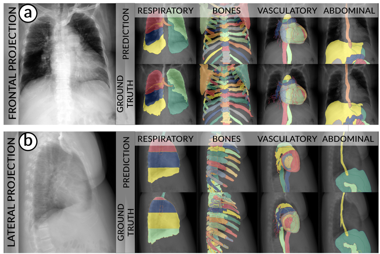 Purpose: Interpreting chest radiographs (CXR) remains challenging due to the ambiguity of overlapping structures such as the lungs, heart, and bones. To address this issue, we propose a novel method for extracting fine-grained anatomical structures in CXR using pseudo-labeling of three-dimensional computed tomography (CT) scans.
Methods: We created a large-scale dataset of 10,021 thoracic CTs with 157 labels and applied an ensemble of 3D anatomy segmentation models to extract anatomical pseudo-labels. These labels were projected onto a two-dimensional plane, similar to the CXR, allowing the training of detailed semantic segmentation models for CXR without any manual annotation effort.
Results: Our resulting segmentation models demonstrated remarkable performance on CXR, with a high average model-annotator agreement between two radiologists with mIoU scores of 0.93 and 0.85 for frontal and lateral anatomy, while inter-annotator agreement remained at 0.95 and 0.83 mIoU. Our anatomical segmentations allowed for the accurate extraction of relevant explainable medical features such as the cardio-thoracic-ratio.
Conclusion: Our method of volumetric pseudo-labeling paired with CT projection offers a promising approach for detailed anatomical segmentation of CXR with a high agreement with human annotators. This technique may have important clinical implications, particularly in the analysis of various thoracic pathologies.
Recommended citation: Seibold, Constantin; Jaus, Alexander; Fink, Matthias A; Kim, Moon; Reiß, Simon; Herrmann, Ken; Kleesiek, Jens; Stiefelhagen, Rainer; . (2023). "Accurate fine-grained segmentation of human anatomy in radiographs via volumetric pseudo-labeling." arXiv preprint arXiv:2306.03934
Download Paper